Frogs in the Classroom

Frogs and biology classrooms have entwined histories. Though frog dissection became an increasingly common practice in secondary and university education during the 20th century, the practical demands of teaching and the often limited availability of live frogs demanded alternate resources.
Posters and models have the advantages of being reusable, predictable, and large enough for sharing by a large class of students. The Whipple Museum possesses many examples of such teaching tools from the late 19th and early 20th centuries, some of which are explored below:
- Our Deyrolle Model reflects the visual and non-textual focus of secondary school biology in 19th century France, and underscores the mimetic purpose of models as alternatives to live frog specimens.
- Teaching tools from the Cambridge Zoology and Comparative Anatomy Department offer a glimpse into the objects used, and even made, by educators here in Cambridge. They embody the visual sensibilities encouraged by anatomical study.
- Biology instruction today has many more resources than those made of plastic, plaster, and paper. Digital resources, which we will call CyberFrogs, reflect a new, digital approach to scientific training that stands in stark contrast to those represented in our collection.
The Department of History and Philosophy of Science is unusual in having teaching programmes connected to the Whipple Museum's world-renowned collection of scientific instruments and books. The Museum is regularly used for both undergraduate and graduate teaching at the University of Cambridge and is a centre of research for students and staff.
Working with the collection
Students are actively encouraged to work with objects in the collection and the undergraduate Part II course includes specific object demonstration classes where students can handle and study the instruments. Lectures and demonstration classes are held in the newly refurbished Reserve Gallery, which provides an ideal space for students to sit down with the objects and interact with them.
Students are also encouraged to produce Case Studies that display their current research or areas of interest in the Whipple's Main Gallery. Other museum displays also complement undergraduate lecture courses, showing instruments and books of the variety described in classroom-taught material.
Starry Messenger
The Whipple Museum developed Starry Messenger, an electronic history of astronomy that focuses on the astronomical instruments and practical uses of the subject. The project was directed by Dr Sachiko Kusukawa and Dr Liba Taub, managed by Dr David Chart and supported by Trinity College, Cambridge.
Starry Messenger draws on the rich collection of instruments and books in the Whipple Collection, the Wren Library and the University Library. It aims to make available online some aspects of the early history of astronomy for student use. The project also provided work experience for the postgraduate students who contributed to its construction.
Excellence in Teaching and Research
For integrating the Whipple Collection into teaching, Professor Liba Taub, Director and Curator of the Museum, was awarded a Pilkington Teaching Prize in 1998. These annual prizes are conferred on academic staff to honour excellence in teaching at the University. The following year, Professor Taub was awarded the Joseph H. Hazen Education Prize by the History of Science Society, in recognition of outstanding contributions to the teaching of history of science. She was particularly commended for her innovative use of museum resources in undergraduate and graduate teaching.
Since 2017, the Board of History and Philosophy of Science has awarded the annual Anita McConnell Prize for an outstanding performance on an essay or dissertation that is based on an object in the Whipple Museum's collection.
Museum classes for Part II students
The Part II course includes lecture-demonstration museum classes on instruments, models and collections. Taking place in the relaxed setting of the Reserve Gallery, the classes allow students to study instruments in the collection relating to their lecture courses, including microscopes, globes and surveying equipment. Students can experience for themselves some of the practical problems faced by contemporary practitioners when using instruments.
The Perse School
The Whipple Museum is partially housed in a large hall with Jacobean hammer-beam roof-trusses, built in 1618 as the first Cambridge Free School. The Main Gallery of the Museum is the original hall of the Perse School, founded as a bequest by Stephen Perse, a fellow of Caius College. The building was completed in 1628 and was described by Nikolaus Pevsner in his 1954 architectural guide to Cambridgeshire as:
"a fine large room with a hammer beam roof. The pendants are as bulbous as Jacobean Bedposts and the spandrels have open work decoration of a scroll type."
The Museum operates on a small budget and relies to a large extent on the generosity of its patrons to support its work. Gifts of all sizes are gratefully received.
Deyrolle's Papier-Mache and Plaster Frog model
The French natural-historical dealership 'Maison Deyrolle' helped link specimen collectors with scientific experts. It also produced scores of models and posters for teaching students. This model of a dissected, pregnant frog is one object within Deyrolle's multimedia, mail-order museum designed to illustrate the wondrous variety of the animal world.
The 'Maison Deyrolle' is a Parisian shop that, since opening in 1831, has dealt in natural history specimens and models, often sourcing items from collectors and selling them to researchers and museums. In the late 19th century it also produced learning materials, which the shop called the 'Musée Scolaire Deyrolle'. These posters and, eventually, models were common materials in French science classes: every school in Paris, Lyon, Bordeaux and Lille used Deyrolle's Musée Scolaire on the recommendation of both the Ministry of Instruction and the Ministry of Agriculture and Commerce.
Most of the 'Musée Scolaire Deyrolle' materials are either posters or larger-than-life models, typically schematic and color-coded. This papier-mâché and plaster frog model presents its subject in a familiar position: on its back, as though under the anatomist's knife, with viscera exposed for easy identification. A clutch of eggs denotes that this is a female frog. Deyrolle's teaching materials were often careful to present their subjects in the same positions in which a student might restrain and dissect their living counterparts. They didn't only represent frogs, but also the ways in which they were actually manipulated and examined by scientists.
Few of the contents of Deyrolle's 'Musée Scolaire' have any labels, unlike the educational material from our own zoology department. Such silence was a deliberate pedagogical choice. Deyrolle considered teaching through images superior to teaching through words: "Visual instruction is the least tiring for the mind, but this education can have good results only if the ideas engraved in the child's mind are rigorously exact." Clarity of form and representational precision were primary values in Deyrolle's commercial world.
Teaching tools from the Cambridge Zoology and Comparative Anatomy Department
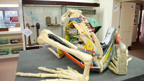
The Whipple Museum's gigantic frog model and anatomical posters were produced for teaching Cambridge students. They show how particular aspects of frogs' bodies would be presented to students by breaking them down into legible, graphic components. Models and posters offered views of frog bodies impossible to achieve through dissection, and are notable more for their abstraction rather than their naturalism.
The Cambridge Department of Zoology and Comparative Anatomy possessed many teaching tools that now reside in the Whipple Museum's collection. Unlike the Ziegler models - renowned for their exquisite craftsmanship and precision - these objects are graphic, simple, and huge. Their construction is guided more by values of clarity and legibility than subtlety and accuracy. We can easily imagine these zoological diagrams displayed before a lecture theatre full of students furiously scribbling notes or snoring softly as their lecturer points out relations between form and function in the frog's cranial nerve.
The monumental wax and plaster model (above) of the frog's skeletal and muscular systems evokes similar teaching contexts. The frog's size and coloration emphasise the rhetorical values of simplicity and clarity, though at the expense of naturalism. Not only are the muscles unrealistically colored, but they are revealed in ways impossible to achieve with a real frog. The species, gender, and age of this specimen are unknown. Students were given small booklets containing the names of each numbered muscle or bone - even those reference booklets specify nothing about what sort of frog we see. It is a perfectly general frog body designed to visualise one specific aspect of its physiology, in this case its musculature. The model was likely produced either bespoke or in-house by the Cambridge Zoology and Comparative Anatomy department in the early 20th century, though no markings identify a maker or date.
Students in Cambridge were asked to produce their own drawings of anatomical forms. These sketches are one student's exemplary drawings in a special notebook distributed to Cambridge students in the 1910s. The author, Professor John Stanley Gardiner, emphasized that students' drawings from life should be "of a semi-diagrammatic nature." If the student completes every page, he wrote, the "Notebook may be made into a Textbook of the Comparative Anatomy of Vertebrates more valuable to its owner than any printed book, both for examination purposes and for subsequent reference."
Cyberfrogs
Frogs have long contributed to teaching anatomy as the source material for schematic representations and as specimens for hands-on anatomical training. That contribution has recently shifted into a new medium: the Internet.
Websites hosting digital frog dissection programmes are almost as old as the Internet itself. The University of Virginia released 'Net-Frog' in 1994. One commentator described it as "truly the first useful Web Site." This site is now defunct, but still accessible via the Internet Archive. More recent programmes include Virtual Frog Dissection, Froguts, and Digital Frog International.
Froguts boasts that its online subscription service includes "Frog, Squid, Starfish, Cow Eye, Owl Pellet, Fetal Pig Modules and The Pea & Fruit Fly Genetics Labs." Digital Frog International touts itself as an improvement on real, physical dissection because the latter lacks built-in reference materials for students: "The Digital Frog 2.5 is so much more effective than a wet lab because the interactive dissection is seamlessly linked to a comprehensive anatomy and physiology section, with human anatomy comparisons. A fascinating ecology section reminds students that biology is the study of living organisms." Digital trips to frogs' different habitats supplement the laboratory dissection module and can be taken with just a click of the mouse.
Schools may subscribe to these services, proponents claim, in order to save money and prevent cruelty to frogs without compromising the quality of their teaching. People for the Ethical Treatment of Animals, a major animal rights organization, argues that, "While dissection may teach some understanding of anatomy, biology, and physiology, it also teaches a profound disrespect for the life it purports to study." Whether students should be obligated to participate in dissections is another matter of controversy. The ethics of animal dissection are always acknowledged as a justification for the very idea of digital dissection. Some scientists and teachers, however, argue that getting one's hands into the frog's real blood and guts is crucial to understanding it, and that digital resources can never replace the real organism in teaching.
Henry Schmidt
Henry Schmidt, 'Frogs in the classroom', Explore Whipple Collections, Whipple Museum of the History of Science, University of Cambridge.
